
iEarLM: A Middle-Outer Ear Lesion Recognition and Clinical Diagnostic Report Generation System Based on Artificial Intelligence and Augmented Reality (AIR) Technology
Category: *Precision Health & Smart Medical
Exhibitor: NATIONAL KAOHSIUNG UNIVERSITY OF SCIENCE AND TECHNOLOGY
Booth No: N416
Characteristic
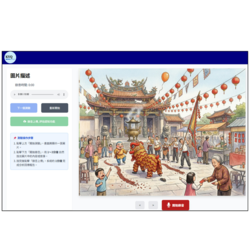
iEarLM: A Middle-Outer Ear Lesion Recognition and Clinical Diagnostic Report Generation System Based on Artificial Intelligence and Augmented Reality (AIR) Technology (hereinafter referred to as the system) consists of a digital otoscope, AR smart glasses, and an edge computing platform, integrated with AI-based image analysis and clinical report generation modules to form a practical intelligent medical assistance system for clinical use. During examination, eardrum images are captured using a digital otoscope and transmitted through a local network to the edge platform for real-time analysis. The system performs lesion localization and contour identification, and the results are simultaneously displayed on both the system interface and AR smart glasses. This enables physicians to directly observe eardrum images and AI analysis results within their field of view, without switching attention between the otoscope and external monitors, thereby facilitating intuitive understanding of lesion location and morphology.
For image analysis, the system adopts an instance segmentation approach to identify eardrum lesions, allowing precise delineation of lesion regions and contours. Compared with existing otoscopic image assistance systems or prior studies that typically provide classification results or bounding-box-based localization, the proposed method further presents lesion shape and spatial extent. This enables image interpretation to evolve from simple abnormality detection to structural understanding of lesions, thereby improving diagnostic consistency and interpretation accuracy. In terms of clinical workflow integration, the system converts recognition outputs into structured medical information and employs a large language model (LLM) with a retrieval-augmented generation (RAG) mechanism to automatically generate draft diagnostic reports. The generated content is grounded in actual recognition results and supplemented by retrieved medical knowledge, ensuring clinical relevance. Physicians are only required to review and refine the draft, enabling completion of clinical documentation within the same workflow. This design integrates image interpretation and report writing, reducing administrative burden and improving operational efficiency.
The system is deployed on an edge computing architecture, allowing both image analysis and report generation to be performed within the hospital environment to ensure real-time performance and data privacy. A functional prototype has been developed, supporting a complete workflow including image acquisition, real-time recognition, visualization, and report generation, and is planned for further clinical validation in real-world settings. The proposed system can be applied in hospital otolaryngology departments, primary care clinics, telemedicine, and remote screening scenarios. In addition, it demonstrates strong potential for integration with digital otoscope devices, medical equipment manufacturers, and healthcare information systems. The clinical data used in this study were approved by the Institutional Review Board (IRB) of Chang Gung Medical Foundation (Approval No. 202500083B0C5001), ensuring that data acquisition and usage comply with medical ethics and privacy regulations, thereby enhancing the reliability of clinical deployment.
Other Products
Products you may be interested in
-
Generative AI care robot-Eirbot
-
YuBelt
-
An integrated microflow cytometry platform-EZ Device
-
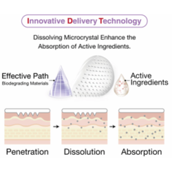
Innovatice Delivery Technology
-
NeuroMirror Neuro-Driven Digital Mirror Rehabilitation System
-
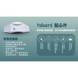
YuGuard
-

Medidata Study Experience
-

Smart Medical Sensing & AI Diagnostic Collaborative Platform
-
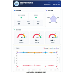
Cognitive Report Page 1
-

2026 KMU IP Portfolio: https://shorturl.at/oFxtr
-
Trianswer
-
The LightRAG and AI Agent architecture
-
Non-Invasive Glucose Monitoring
-
COMET
-
AI-MindSnap
-
Endourology & Complete Urinary System Simulation Platform
-
Labyrinth One-Automated CTC Enrichment System
-

Gentle Plasma Skin Regenerator Model 2 Plus
-
Metastable β Titanium-Rich Medium-Entropy Alloy and Preparation Method
-
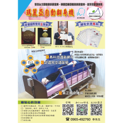
Maria Automatic Turning Bed
-

iHealing Future: Intelligent Wound Sensing System
-
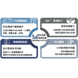
Virtual Reality Pediatric Emergency Skills Training and Clinical Response Assessment System
-

Medidata Patient Experience
-
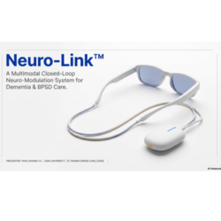
Neuro-Link Smart Dementia Care Neuromodulation System
-
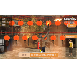
Leisure-Based Virtual Reality Physical and Cognitive Training System: Jiufen Edition
Highest Rated Products
-
Pneumatic Liquid Filling Machine
-
Powder Machine-High Speed Mixer / Granulator
-
Planetary Vacuum Mixer
-
SEMI-AUTOMATIC EYESHADOW PRESS MACHINE
-
2ton Mixing and Melting Tank with Storage Tanks
-
High Speed Mixing Machine
-
In-Line Homogenizer
-
VACUUM EMULSIFY MIXING EQUIPMENT (Bottom Homogenized Series)
-
VACUUM EMULSIFY MIXING EQUIPMENT
-
VACUUM EMULSIFY MIXING EQUIPMENT
-
UV-Visible Spectrophotometer
-
Cell Function Assay Kit(Apoptosis/Cycle/Proliferation/Cytotoxicity/Viability)
-
CM30 Incubation Monitoring System
-
CURIOSIS Celloger Nano
-
DISCOVERY Comfort Pipettor
-
LTS Style Pipette Tips
-
Rotary Evaporator
-

Centrifugal Evaporator (Teflon coated)
-
Clinical Small Centrifuges
-
High Performance Liquid Chromatography, HPLC
-
WINMAPᵀᴹ
-
DS MAPs
-
Vaccine Microarray Patches
-
MiniMAP™
-
Power Patch
-

V Patch
-

Schrodinger
-
SuitNifty Scar Gel (Non-Sterile)
-

Natural Marine Collagen
-
TISUCODETM Type II Collagen Capsules


